AAV2 VP1 + VP2 + VP3, recombinant proteins, set
View more

View more

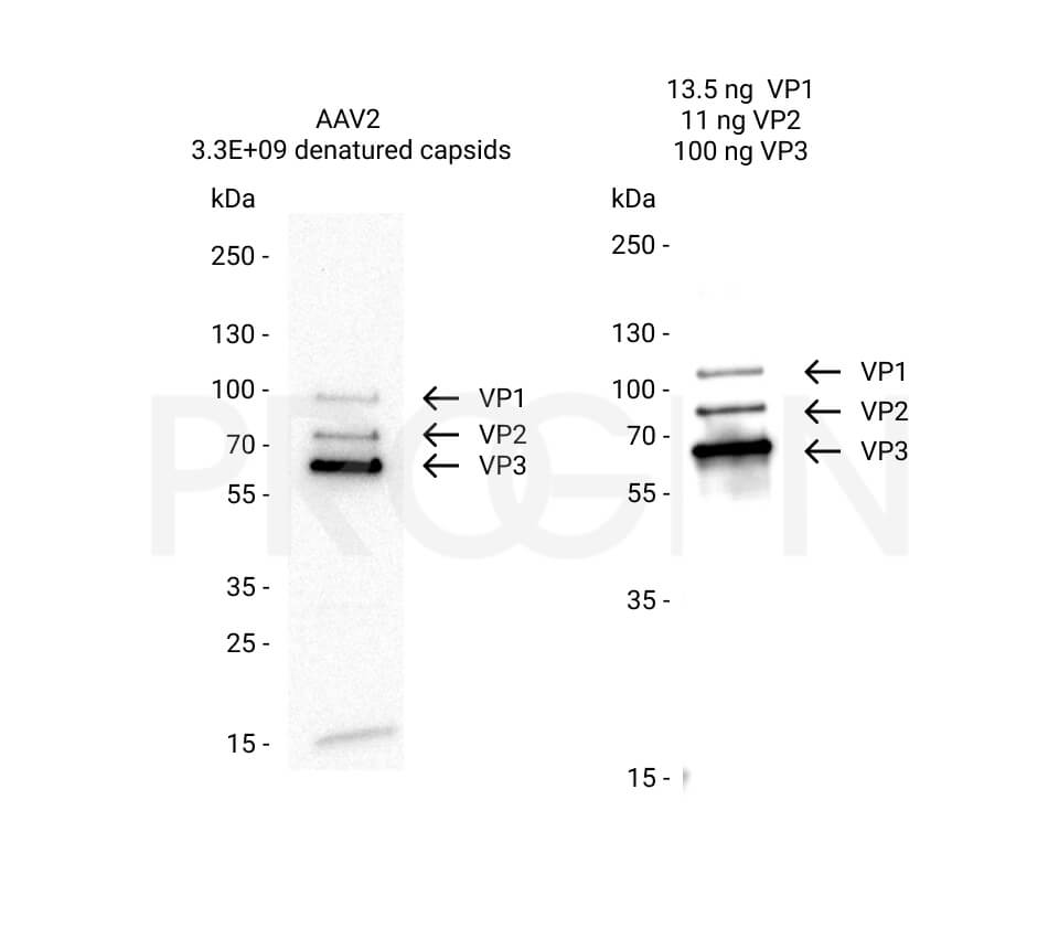
View more

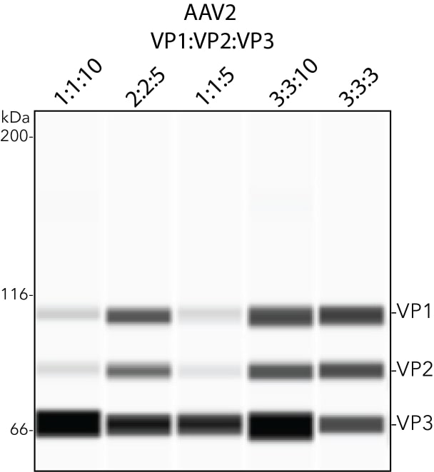
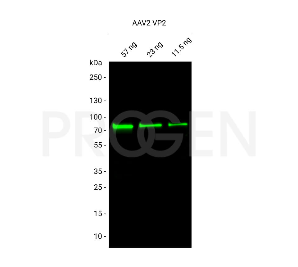
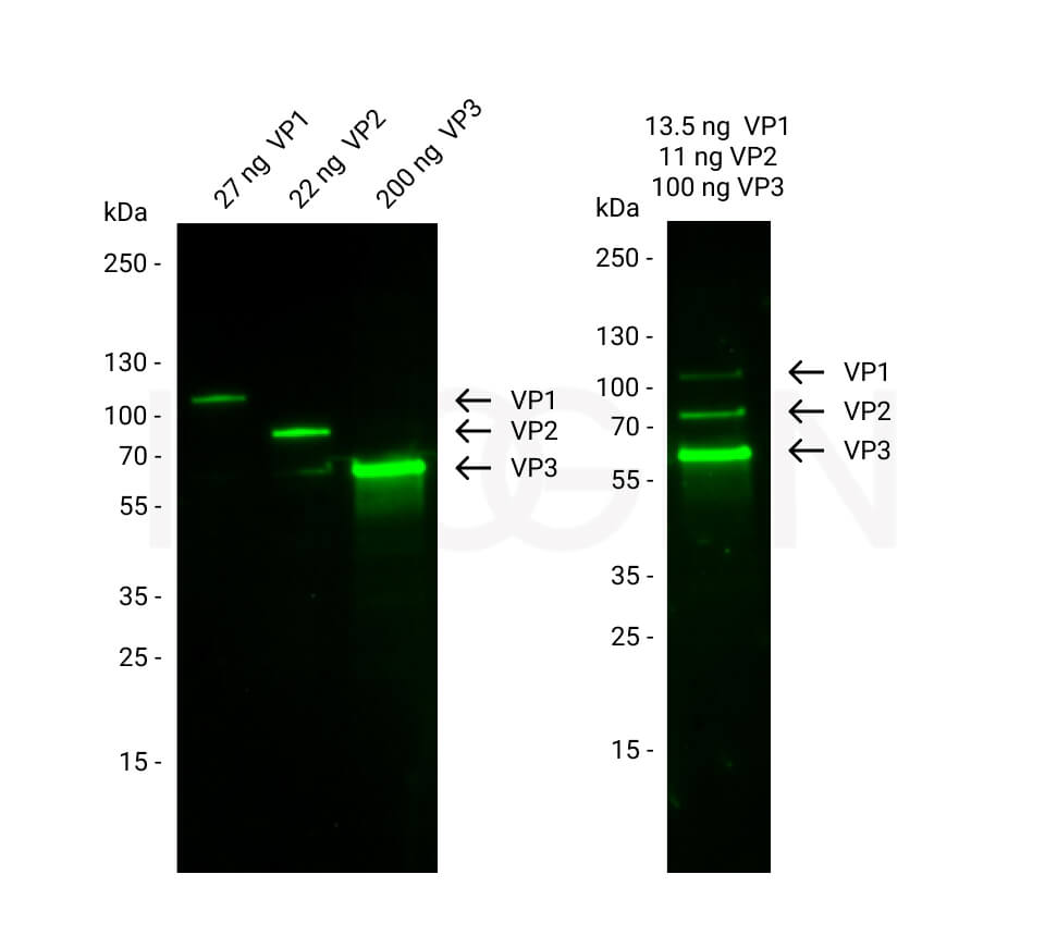
Analysis of AAV2 VP1 + VP2 + VP3, recombinant proteins (Cat. No. 72001) with various stoichiometric ratios by Simple WesternTM, a CE-immunoassay platform from ProteinSimple, a Bio-Techne brand. Capsid proteins were detected using an anti-AAV VP1/VP2/VP3 mouse monoclonal antibody (Cat. No. 690058).
View more
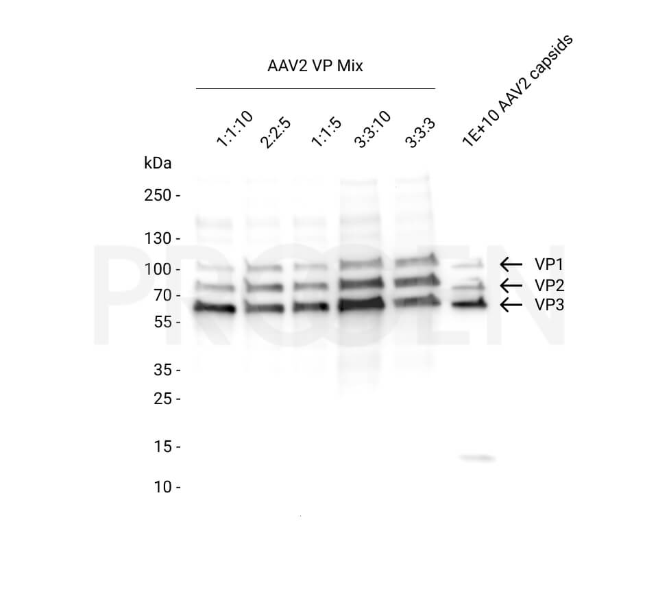
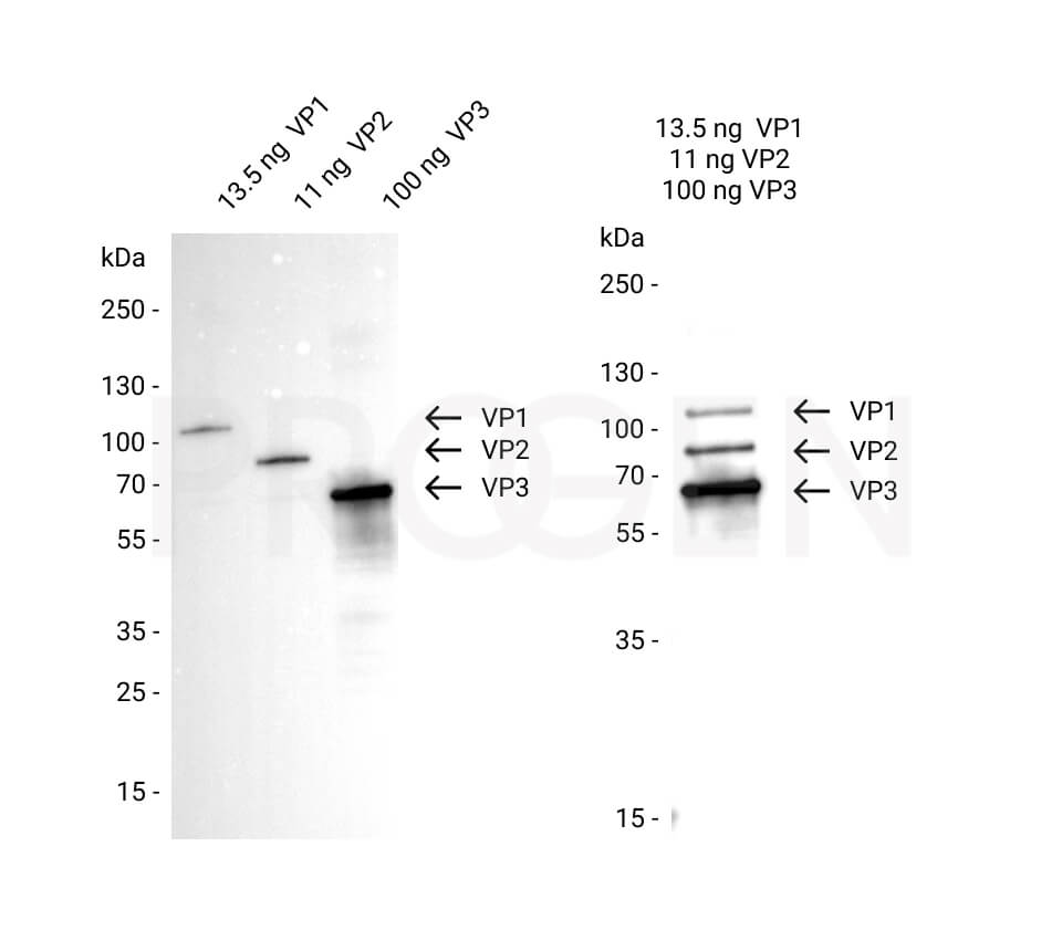
Western blot analysis was performed on different molar ratios of VP1:VP2:VP3 (1:1:10, 2:2:5, 1:1:5, 3:3:10 and 3:3:3) compared to 1E+10 denatured AAV2 capsids. The capsids were denatured for 10 min at 95°C. The PVDF membrane was blocked with 5% dry milk in PBST (PBS + 0.1% Tween 20) for 1 h at RT. The primary antibody anti-AAV VP1/VP2/VP3 mouse monoclonal, B1 (Cat. No. 690058) was diluted in blocking buffer (antibody concentration 0.5 µg/ml) and incubated for 1 h at RT. The secondary antibody goat anti-mouse IgG HRP was also diluted in blocking buffer (antibody concentration 0.2 µg/ml) and incubated for 1 h at RT. The bands were visualized by chemiluminescent detection using PierceTM ECL Western Blotting Substrate.
Please note that various amounts of total protein were loaded in each lane.
View more

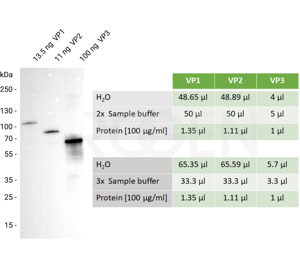
10 µl of each solution can be separately loaded onto the SDS PAGE and analyzed by Western blot using the B1 antibody (Cat. No. 690058, Cat. No. 61058-488, Cat. No. 61058-647).
View more

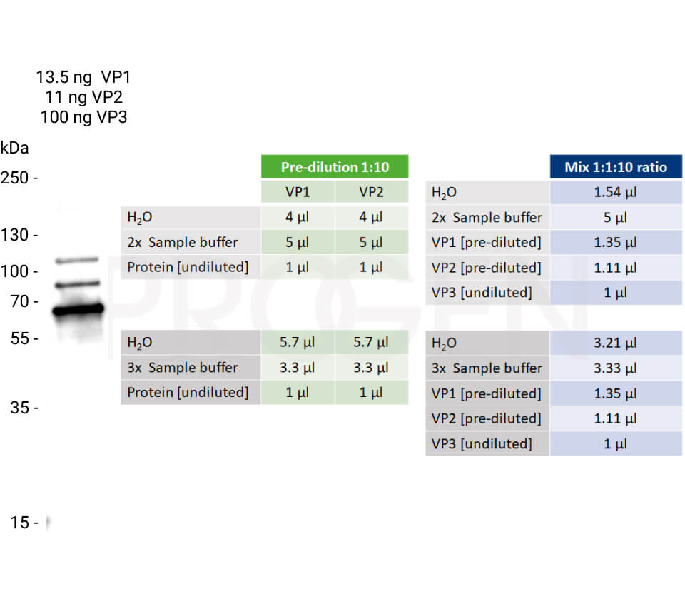
Undiluted = 100 µg/ml, pre-diluted = 10 µg/ml
View more

View more



Analysis of AAV2 VP1 + VP2 + VP3, recombinant proteins (Cat. No. 72001) with various stoichiometric ratios by Simple WesternTM, a CE-immunoassay platform from ProteinSimple, a Bio-Techne brand. Capsid proteins were detected using an anti-AAV VP1/VP2/VP3 mouse monoclonal antibody (Cat. No. 690058).


Western blot analysis was performed on different molar ratios of VP1:VP2:VP3 (1:1:10, 2:2:5, 1:1:5, 3:3:10 and 3:3:3) compared to 1E+10 denatured AAV2 capsids. The capsids were denatured for 10 min at 95°C. The PVDF membrane was blocked with 5% dry milk in PBST (PBS + 0.1% Tween 20) for 1 h at RT. The primary antibody anti-AAV VP1/VP2/VP3 mouse monoclonal, B1 (Cat. No. 690058) was diluted in blocking buffer (antibody concentration 0.5 µg/ml) and incubated for 1 h at RT. The secondary antibody goat anti-mouse IgG HRP was also diluted in blocking buffer (antibody concentration 0.2 µg/ml) and incubated for 1 h at RT. The bands were visualized by chemiluminescent detection using PierceTM ECL Western Blotting Substrate.
Please note that various amounts of total protein were loaded in each lane.


10 µl of each solution can be separately loaded onto the SDS PAGE and analyzed by Western blot using the B1 antibody (Cat. No. 690058, Cat. No. 61058-488, Cat. No. 61058-647).


Undiluted = 100 µg/ml, pre-diluted = 10 µg/ml


• Useful for ratio analysis
• Precise molar concentration
• Suitable for WB, dot blot, CE, SDS PAGE
Included products:
Product description
| Quantity | 10 µg each protein |
|---|---|
| Application | Capillary electrophoresis (CE), Dot blot, SDS PAGE, WB |
| Purification | Ni-NTA chromatography |
| Storage | -80°C |
| Intended use | Research use only |
| Concentration | 100 µg/ml (VP1: 1.19 µM, VP2: 1.45 µM, VP3: 1.61 µM) |
| Formulation | Liquid, 6 M urea in PBS |
| Source | Escherichia coli |
| Molecular weight | VP1: 84.1 kDa, VP2: 68.9 kDa, VP3: 62.2 kDa (calculated Mw from aa sequence) |
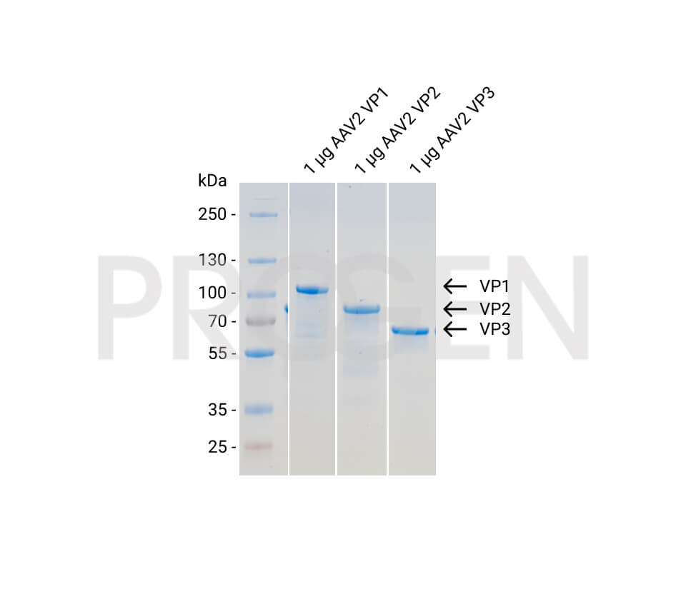
| Purity | > 90% (determined by SDS PAGE) |
| Product description | N-terminal His-tagged (MGSSHHHHHHSSGLVPRGSH) recombinant AAV2 capsid proteins VP1 + VP2 + VP3 |
Applications
| Tested applications | Tested dilutions |
|---|---|
| Capillary electrophoresis (CE) | Assay dependent |
| Dot Blot | 100 ng, depending on primary antibody and detection method |
| SDS PAGE | 1 µg |
| Western Blot (WB) | 5-20 ng, depending on primary antibody and detection method |
Background
The AAV capsid consists of three capsid proteins, i.e. VP1, VP2 and VP3, which differ in their N-terminus and encapsulate the genomic ssDNA. In native virus particles, the three proteins form subunits with a ratio of 1:1:10 (VP1:VP2:VP3), in a total number of 60 subunits per capsid.
This set of recombinant AAV2 VP1, VP2 and VP3 can be used to create a mixture with the precise molar ratio of 1:1:10 to compare the protein composition of the viral capsid in your sample by protein detection methods, e.g. western blot.
All three recombinant AAV2 capsid proteins are available as set (Cat. No. 72001) or as individual proteins (Cat. No. 640823, 640824, 640825).
Downloads
Q & A's
Customer Reviews
0 of 0 reviews
Login
Progen Support
For scientific and customer support, you can reach us by phone or email.
Related Products
- Consists of fully assembled, empty AAV2 capsids
- Represents serotype-specific conformational epitopes
- Reliable positive control for ELISA
- Fully assembled, empty AAV2 capsids
- Serotype-specific conformational epitopes
- Reliable positive control for dot blot, western blot, ELISA
- Quality controlled for purity, AAV titer and filling grade
- Alignment to internal reference standard material
For US shipment, the packages are only sent out for delivery on Mondays and Tuesdays, in the EU from Monday until Wednesday.
- Fully assembled AAV2 capsids filled with an eGFP reporter gene controlled by a CMV promoter
- Serotype-specific conformational epitopes
- Reliable positive control for dot blot, western blot, ELISA, PCR and cell-based assays
- Quality controlled for purity, filling grade, aggregation, total capsid and genome titer
For US shipment, the packages are only sent out for delivery on Mondays and Tuesdays, in the EU from Monday until Wednesday.
- ELISA for quantitation of AAV2 capsids
- Detection of full and empty AAV2 capsids
- A20 antibody is used as capture and detection antibody
- ELISA for quantitation of AAV2 capsids
- Detection of full and empty AAV2 capsids
- A20R antibody is used as capture and detection antibody
• Useful for ratio analysis
• Precise molar concentration
• Suitable for WB, dot blot, CE, SDS PAGE
• Useful in combination with AAV2 VP2 and VP3 for ratio analysis (also available as set, Cat. No. 72001)
• Precise molar concentration
• Suitable for WB, dot blot, CE, SDS PAGE
• Useful in combination with AAV2 VP1 and VP3 for ratio analysis (also available as set, Cat. No. 72001)
• Precise molar concentration
• Suitable for WB, dot blot, CE, SDS PAGE
• Useful in combination with AAV2 VP1 and VP2 for ratio analysis (also available as set, Cat. No. 72001)
• Precise molar concentration
• Suitable for WB, dot blot, CE, SDS PAGE
- get results in less than 2 hours and increase your daily sample throughput
- easy workflow & process integration
- same accuracy and reproducibility as the standard AAV2 Titration ELISAs
- A20R antibody is used as capture and detection antibody
- Mouse monoclonal
- Suitable for ELISA, ICC/IF, IP and WB
- Reacts with VP1 of AAV1, AAV2, AAV3, AAV5, AAV6, AAV7, AAV8, AAV9 and AAVDJ
- Isotype: IgG2a
- Mouse monoclonal
- Suitable for ICC/IF, IP and WB
- Reacts with AAV2 and AAVDJ
- Isotype: IgG1
- Mouse monoclonal
- Suitable for affinity chromatography, dot blot, ICC/IF, IP and WB
- Reacts with denatured VP1, VP2, VP3 proteins of AAV1, 2, 3, 5, 6, 7, 8, 9, rh10, DJ
- Isotype: IgG1
- Rabbit polyclonal
- Suitable for ELISA, ICC/IF, IP and WB
- Reacts with AAV1, AAV2, AAV3, AAV5, AAV6, AAV7, AAV8, AAV9, AAVrh10, AAVDJ and AAVrh74, weakly with AAV4
- Strong affinity for denatured capsid proteins
- Weak affinity for native capsids
- Purified, lyophilized
- Mouse monoclonal
- Suitable for dot blot, ELISA, ICC and neutralization assay
- Reacts with AAV2, AAV2 7m8 and AAV3 intact particles
- Isotype: IgG3
- Mouse monoclonal
- Suitable for Affinity Chromatography, ICC/IF and IP
- Reacts with AAV2 Rep40, Rep52, Rep68 and Rep78
- Isotype IgG1
- Purified, lyophilized
- Mouse monoclonal
- Suitable for WB
- Reacts with AAV2 Rep40, Rep52, Rep68 and Rep78
- Isotype: IgG1
- Mouse monoclonal
- Suitable for WB
- Reacts with AAV2 Rep40, Rep52, Rep68 and Rep78
- Isotype: IgG1
- Mouse Monoclonal
- Suitable for WB, ICC/IF and IP
- Reacts with AAV2 Rep52 and Rep78
- Isotype: IgG1
- Recombinant chimeric (human Fc region)
- Suitable for dot blot, neutralization assay and serology ELISA
- Reacts with AAV2 and AAV3 intact capsids
- Isotype: IgG1
- Rapid lateral flow test for the detection of AAV2 and AAV3 capsids
- Semi-quantitative AAV titer determination in cell lysates and purified preparations
- Detection of full and empty AAV2 and AAV3 capsids
- Highly effective density gradient media for the separation of full and empty AAV particles
- Designed for the purification of viruses (such as AAV)
- Useful to isolate different cell types and organelles
- Ready-made, sterile and endotoxin-tested solution of iodixanol
- Automated ELISA kit for quantification of AAV2 virons and assembled capsids
- A20R antibody (Cat. No. 610298) is used as capture and detection antibody
- Suitable for the ProteinSimple Ella system


