The perilipin/PAT family

tackle lipid metabolism


Perilipins, also called lipid droplet coating proteins or PLINs, are phosphoproteins encoded by the PLIN gene. They are located at the surface of lipid droplets (LDs), which store energy in lipids. These droplets are cellular organelles mostly found in adipocytes, cells that compose the adipose tissue. The lipid droplet consists of a core of triacylglycerols and cholesterinesters, surrounded by a phospholipid and cholesterol monolayer with associated coating proteins, the most abundant being perlipins (PAT family). These perilipins regulate the lipid metabolism and storage in adipocytes, protecting against lipolysis by natural occurring lipases. Furthermore they have a structural role and possibly contribute to the formation of lipid droplets. Another point worth noting is that important lipases like the hormone-sensitive lipase (HSL) or the adipose triglyceride lipase (ADGL) are able to travel to the perilipin scaffold and assemble the lipolytic complex by binding to phosphorylated perilipins. As a consequence lipolysis can occur.

Because LDs are linked to several diseases like diabetes, obesity, liposarcoma,  atherosclerosis, lipid droplet biogenesis and several viral or bacterial infections, it is quite important to understand and characterize Perilipins. 


Browse Perilipin Antibodies

"In the community of lipid droplet biology research, your antibodies are highly appreciated and many papers in this field rely on PROGEN antibodies."

Prof. Dr. Matthijs Hesselink, Maastricht University Medical Center, The Netherlands

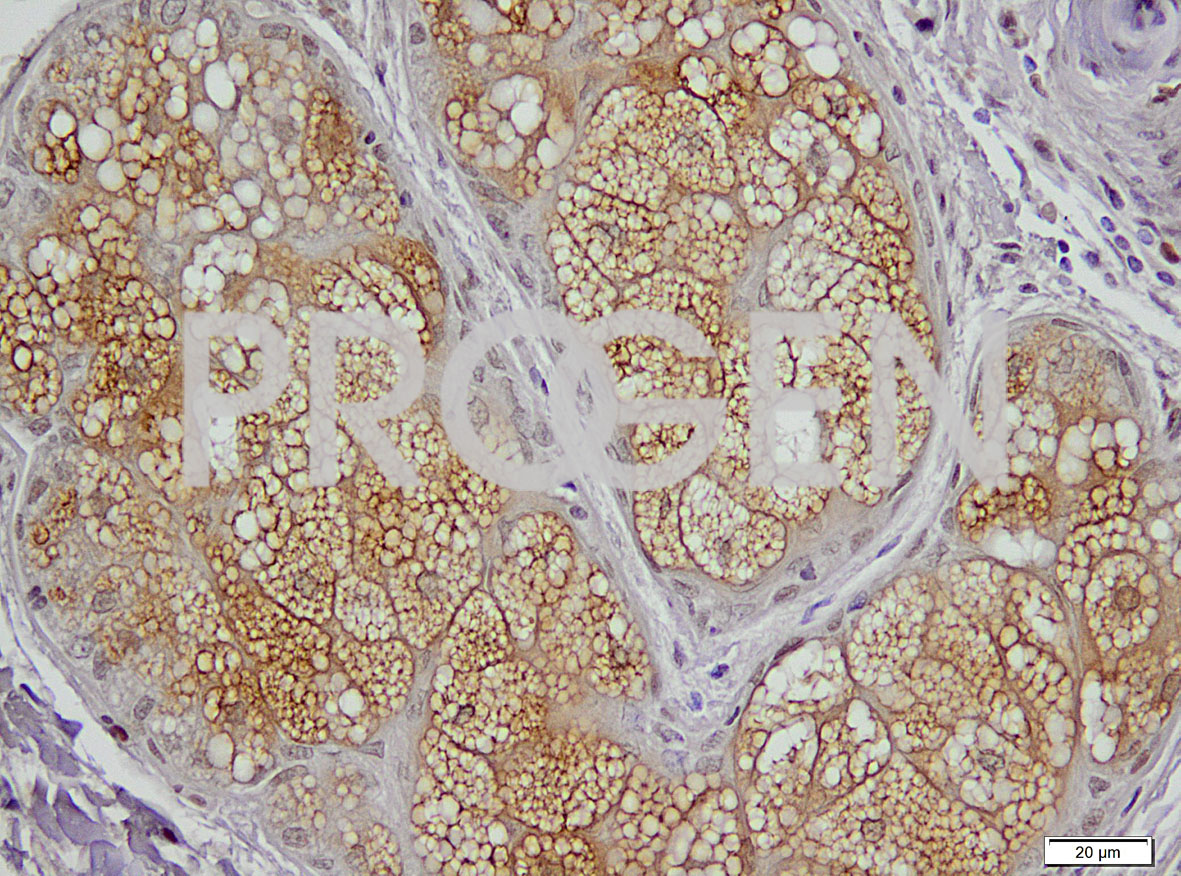
Perilipin-2 immunohistochemistry of human skin; © PROGEN in cooperation with Uniklinik Heidelberg, Dr. Jochen Heß

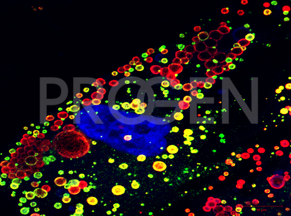
Immunofluorescent double-staining of perilipin-1 (red) and perilipin-2 (green) in human preadipose cells, DAPI-labelled nucleus (purple); © W. Franke, DKFZ Heidelberg

Research antibodies

Sit amet, consetetur sadipscing elitr

In Mammals there are five major perilipins (perilipin 1-5). Lipid droplets in other cell types contain only PLIN 2 and 3 at the surface. Depending on the lipid droplet composition different perilipins are present. All Perilipin-Proteins share the PAT domain but have unique C-termini. Some also have alternate splice versions, wich leads to even more variety within the family. Perilipin 1A, 2 and 5 are present predominantly on the surface of lipid droplets rich in triacylglycerols. If the droplet is enriched in cholesterol esters perilipins 1C, 1D and 4 are preferentially bound. Usually PLIN 2 and 3 are ubiquitionelly expressed whereas PLIN 1,4 and 5 habe more limited tissue expression, due to their function as lipase-controlling proteins.

PROGEN offers a selecion of different antibodies against all 5 perilipin subtypes (PLIN1-PLIN5) for a number of applications (western blot, IHC, IF and ICC).


  • Other names: Perilipin A, B, C, D (without Number)
  • Typically on the surfaces of intracellular lipid storage droplets
  • Additional splice variants 1A 1B 1C 1D, Major isoform: 1A, shorter isoforms: 1C and 1D
  • Adrenal gland, adipocytes of white/brown adipose tissue, cell culture e.g. 3T3-L1 adipocytes,steroidogenic adrenal cortical, Leydig cells
  • Useful pathological marker (de novo expressed in hepatocyte steatogenesis)

View PLIN1 Antibodies

  • Other names: adipocyte differentiation-related protein (ADRP) / ADFP / adipophilin
  • Tightly associated with the LD surface, typically referred to smaller LDs
  • Relativly permissive to lypolyse
  • Enhanced expression as marker for pathologies characterized by increased lipid droplet accumulation e.g. atheroma, steatosis, obesity and certain cases of liposarcoma
  • Considered to be a potent marker for atherosclerosis

View PLIN2 Antibodies

  • Other names: TIP47 / placental protein 17 (PP17) / M6PR binding protein 1 (MP6PRBP1)
  • Sequence similarity to PLIN2
  • Tightly associated with the LD surface, typically referred to smaller LDs
  • Suggested to contribute to LD formation and stabilzation.

View PLIN3 Antibodies

  • Other names: S3-12
  • Selectively expressed in adipocytes in the middle to late stage of differentiation
  • Might be involved in uptake of lipids and hydrophobic substances

View PLIN4 Antibodies

  • Other names: OXPAT / MDLP
  • Functionally associated with lipid storage droplets
  • Found in muscle cell tissue
  • Supposed to be involved in the supply of energy to the mitochondria of muscle cells
  • Faciliates FA oxidation in skeletal muscle on demand

View PLIN5 Antibodies

Publications

  1. Heid, H. et al. Lipid droplets, perilipins and cytokeratins--unravelled liaisons in epithelium-derived cells. PLoS One 8, (2013).
  2. Fernández-Rojo, M. A. et al. Caveolin-1 Is Necessary for Hepatic Oxidative Lipid Metabolism: Evidence for Crosstalk between Caveolin-1 and Bile Acid Signaling. Cell Rep. 4, 238–247 (2013).
  3. Kuramoto, K. et al. Deficiency of a Lipid Droplet Protein, Perilipin 5, Suppresses Myocardial Lipid Accumulation, Thereby Preventing Type 1 Diabetes- Induced Heart Malfunction. Mol. Cell. Biol. 34, 2721–2731 (2014).
  4. Barquissau, V. et al. White-to-brite conversion in human adipocytes promotes metabolic reprogramming towards fatty acid anabolic and catabolic pathways. Mol. Metab. 5, 352–366 (2016).
  5. Gallardo-Montejano, V. I. et al. Nuclear Perilipin 5 integrates lipid droplet lipolysis with PGC-1a/SIRT1-dependent transcriptional regulation of mitochondrial function. Nat. Commun. 7, (2016).
  6. Inoue, J. et al. Identification of BCL11B as a regulator of adipogenesis. Sci. Rep. 6, 32750 (2016).
  7. Kaushik, S. & Cuervo, A. M. AMPK-dependent phosphorylation of lipid droplet protein PLIN2 triggers its degradation by CMA. Autophagy 12, 432–438 (2016).
  8. Furukawa, S., Nagaike, M. & Ozaki, K. Databases for technical aspects of immunohistochemistry. J. Toxicol. Pathol. 30, 79–107 (2017).
  9. Gemmink, A. et al. Dissociation of intramyocellular lipid storage and insulin resistance in trained athletes and type 2 diabetes patients; involvement of perilipin 5? J. Physiol. (2017).
  10. Morales P.,Bucarey J. Espinosa A. Muscle Lipid Metabolism: Role of Lipid Droplets and Perilipins J Diabetes Res. (2017)
  11. Sztalryd C., Brasaemle D. The perilipin family of lipid droplet proteins: Gatekeepers of intracellular lipolysis. Biochim Biophys Acta Mol Cell Biol Lipids (2017)
  12. Bickel P., Tansey J., Welte M. PAT proteins, an ancient family of lipid droplet proteins that regulate cellular lipid stores Biochim Biophys Acta. 2009 Jun; 1791(6): 419–440. (2009)

Know what you need?

Login and order directly
from your account.

Order Now

Need more information?

Visit our support hub for FAQs,
references and downloads.

Visit Support Hub