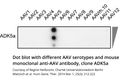
Binding Sites and Reactivity
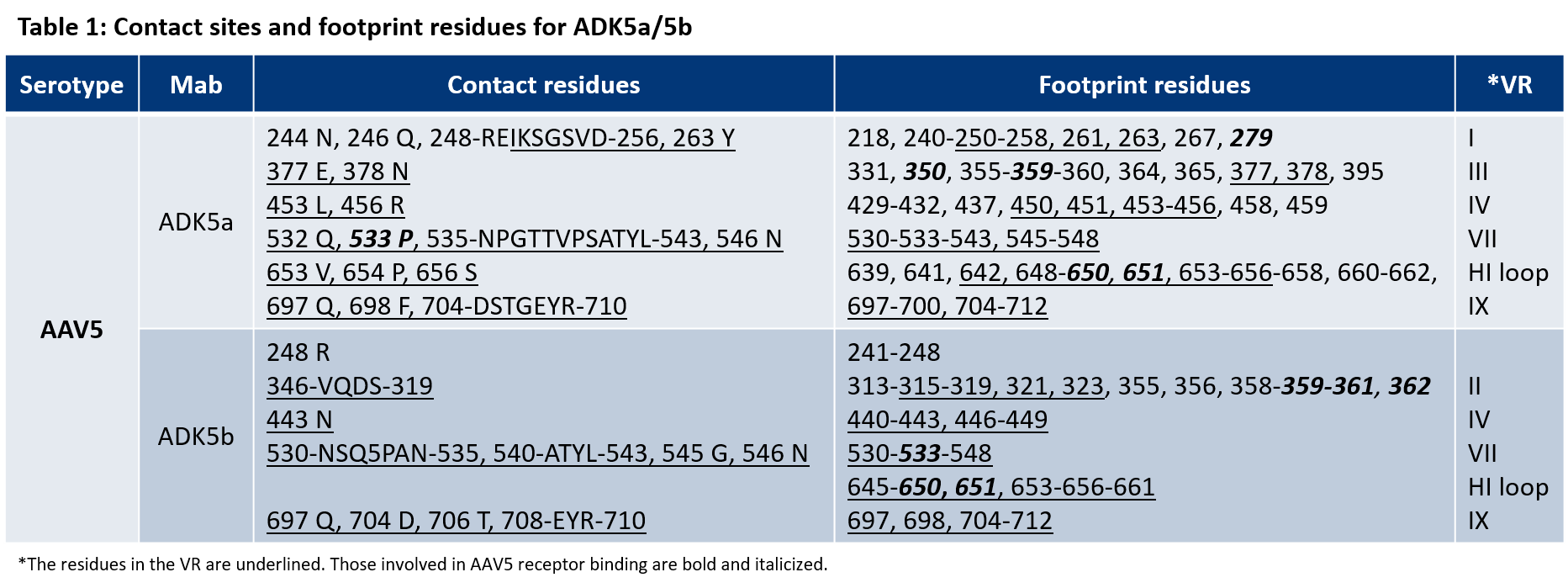
The ADK5a antibody used for the AAV5 Titration ELISA as well as the ADK5b specifically recognize assembled AAV5 capsids, hence epitopes that result from a specific conformational assembly of the capsid proteins VP1, VP2 and VP3. In the publication cited below the contact sites and footprint residues identified for ADK5a and ADK5b are described and the results are summarized in table 1.
Multiple contact sites and footprint residues have been identified, that are very likely to be part of the binding site. The amino acids of each binding site are located in different parts of the protein chains and are recognized as the epitope of the antibody only in the assembled capsid where they are in close proximity to each other and in the correct conformation.
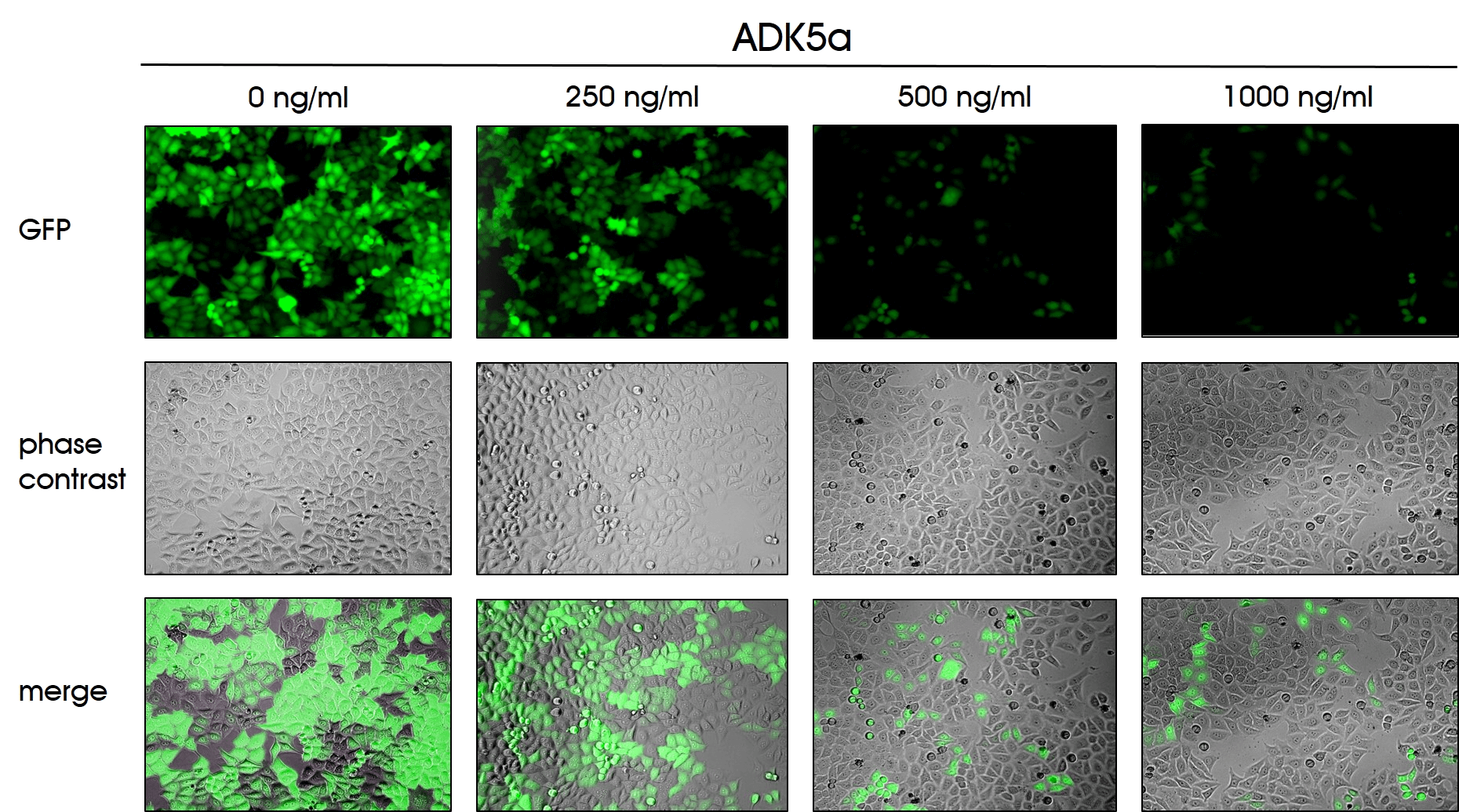
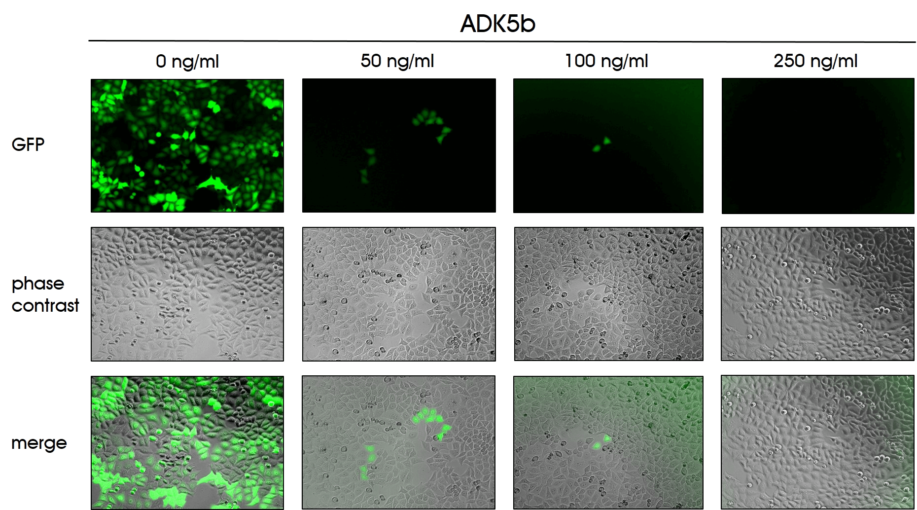
The ADK5a (Cat. No. 610148) and the ADK5b antibody (Cat. No. 610149) were tested for neutralization using different antibody concentrations. After the HeLa cells were seeded in a 96-well plate they were incubated for 48h with the AAV5 particles containing a GFP reporter and the indicated concentration of the ADK5a or ADK5b antibody, respectively.
The microscopic analysis of the cells clearly demonstrates the neutralizing activity of the ADK5a or ADK5b antibody by decreasing GFP expression with increasing antibody concentration.
Available Products
 and secondary anti-rabbit HRP antibody (Cat. No. 90003, 1:10,000), sample AAV5 capsids (75-400 ng)")
- Rabbit polyclonal
- Suitable for Affinity Chromatography and WB
- Reacts with AAV5
 mouse monoclonal, ADK5a, lyophilized, purified")
- Purified, lyophilized
- Mouse monoclonal
- Suitable for dot blot, ELISA and neutralization assay
- Reacts with AAV5 intact capsids
- Isotype: IgG2a kappa
 as detection antibody (1:30)")
- Biotin conjugate
- Mouse monoclonal
- Suitable for ELISA
- Reacts with AAV5 intact capsids
- Isotype: IgG2a kappa
 mouse monoclonal, ADK5b, lyophilized, purified")
- Purified, lyophilized
- Mouse monoclonal
- Suitable for ELISA, ICC and neutralization assays
- Reacts with AAV5 intact capsids
- Isotype: IgG2b kappa