AAV Capsid Protein Antibodies

Detect viral capsid proteins VP1, VP2 and VP3
PROGEN's AAV capsid protein antibodies recognize linear epitopes of the viral capsid proteins VP1, VP2 and VP3. The high sequence similiarity of AAV capsid proteins allows the easy detection of proteins across serotypes.
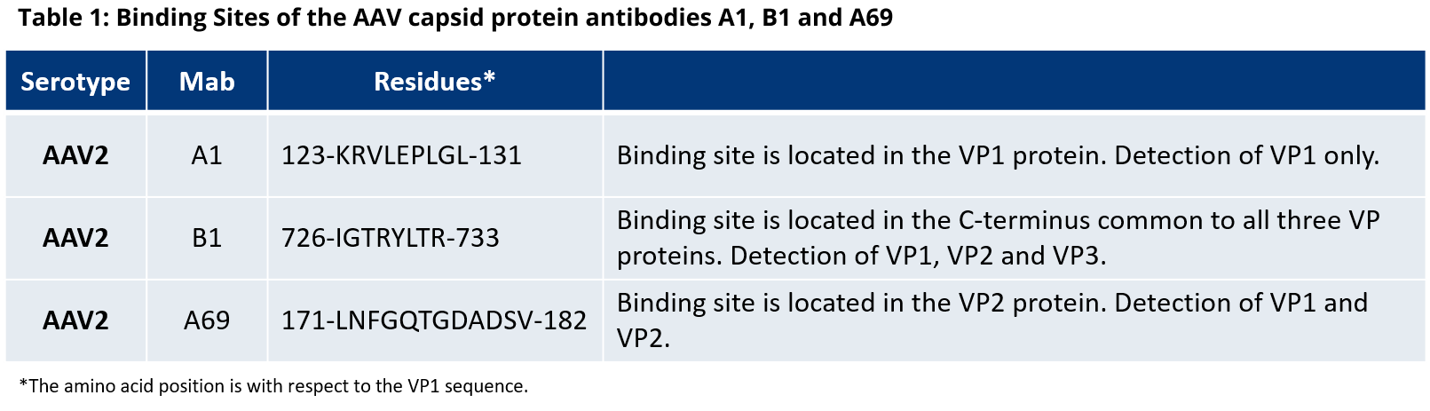
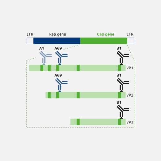
Figure 1 shows the three capsid proteins and the corresponding antibody binding sites. As all VP proteins are encoded by the same reading frame they only differ in their N-terminal region due to the use of an alternate start codon and alternate splicing. Antibodies binding the C-terminal part, e.g. B1, are able to bind to all of the capsid proteins whereas antibodies like A1 can only bind to VP1 and A69 to VP1 and VP2.
Capsid protein antibodies can be used in western blot analyses to detect the expression levels of VP1, VP2 and VP3, as well as for protein localization studies or protein-protein interactions using chromatography, immunoprecipitation or immunofluorescence.
Binding Sites and Reactivity
Capsid protein antibodies were raised in mice against AAV2 capsid proteins and are suitable for all applications detecting linear epitopes of the denatured VP protein, such as western blot analysis.
Cross-reactivity
A1
Reacts with VP1 proteins from all common serotypes AAV1-12. In immunoprecipitation a reaction with a non-AAV derived protein can occasionally be seen.
B1
The B1 antibody shows high cross-reactivity with most of the tested AAV serotypes due to high sequence similarities of VP proteins derived from different AAVs.
Using AFDyes
Using an AFDye™ labeled antibody allows you to detect your proteins of interest without using a secondary antibody. This saves you valuable time as you can also simultaneously detect different proteins in the same sample by using differently labelled antibodies e.g. AFDye™ 647 and AFDye™ 488.
PROGEN offers two different AFDye™ conjugates for the B1 antibody: AFDye™ 647 and AFDye™ 488.
AFDyeTM 647
This western blot shows the analysis of different AAV serotpyes using our anti-AAV VP1/VP2/VP3 AFDyeTM 647 antibody.

AFDyeTM 488
This western blot shows the analysis of different AAV serotypes using our anti-AAV VP1/VP2/VP3 AFDyeTM 488 antibody.

AAV Antibodies FAQs
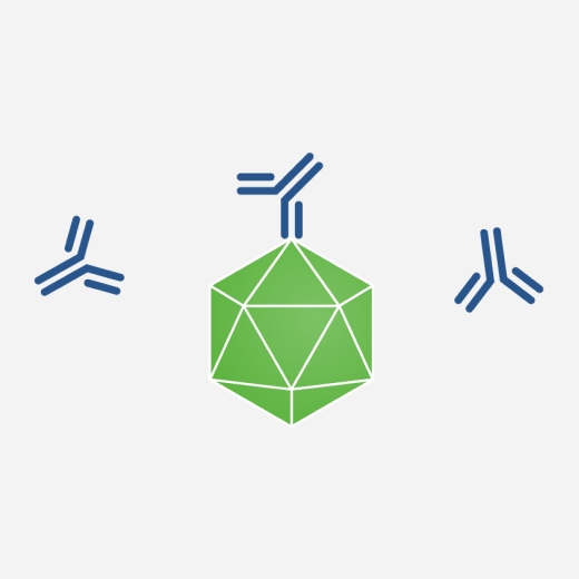
Yes, our AAV particle antibodies detect conformational epitopes only present on assembled capsids.
You can find all AAV particle antibodies here.
In contrast, AAV capsid protein antibodies recognize linear epitopes present on denatured VP proteins.
You can find all AAV capsid protein antibodies here.
Yes, PROGEN offers a selection of AAV antibodies in 10 µg sample sizes. You can find all available AAV antibodies here: AAV Antibodies
Anti-Adeno-Associated Virus Capsid Proteins Antibodies
Anti-Adeno-Associated Virus Particle Antibodies
For more information on the antibody binding epitopes on assembled AAV1, 2, 5, 8 and 9 capsids, see the tables below summarizing the published data from the following publications on the specific antibody binding sites: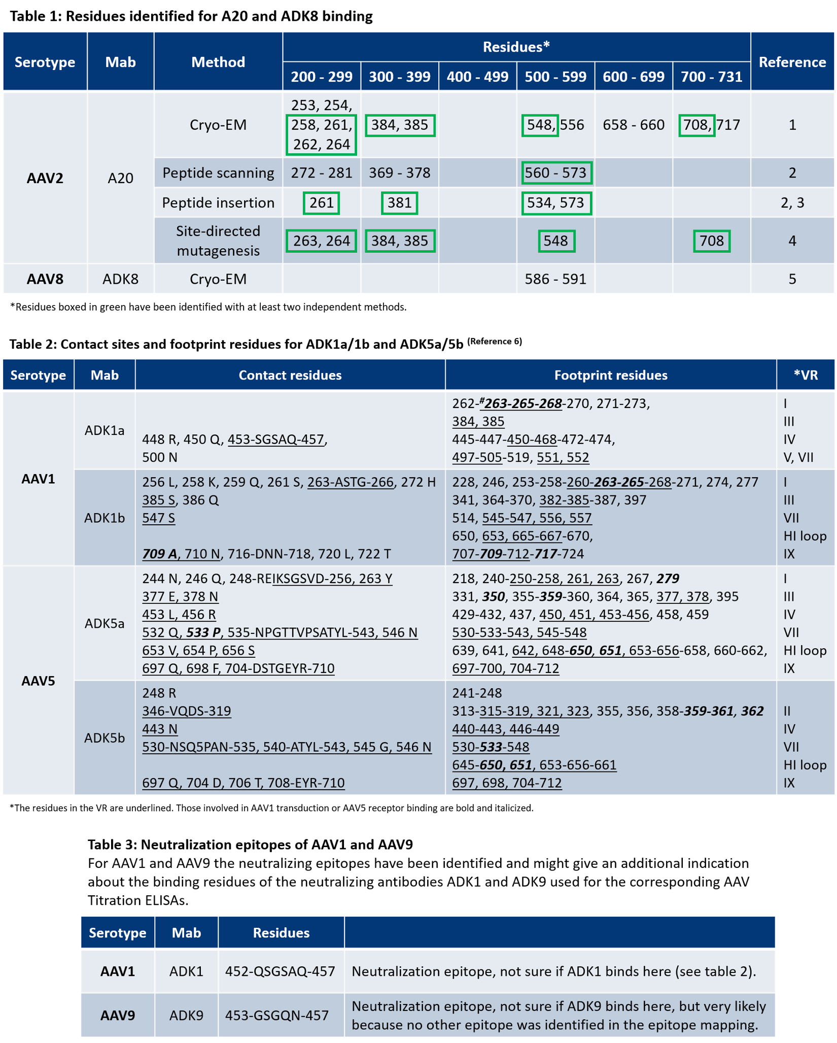
-
AAV 1
- Tseng, Y.-S. et al. Adeno-Associated Virus Serotype 1 (AAV1)-and AAV5-Antibody Complex Structures Reveal Evolutionary Commonalities in Parvovirus Antigenic Reactivity. J. Virol. 89, 1794–1808 (2015).
- McCraw et al. Structureof adeno-associated virus-2 in complex with neutralizing monoclonal antibody A20. Virology (2012) 431:40-9.
- Wobus, C. E. et al. Monoclonal antibodies against the adeno-associated virus type 2 (AAV-2) capsid: epitope mapping and identification of capsid domains involved in AAV-2-cell interaction and neutralization of AAV-2 infection. J. Virol. 74, 9281–93 (2000).
- Huttner et al. Genetic modifications of the adeno-associated virus type 2 capsid reduce the affinity and the neutralizing effects of human serum antibodies. Gene Ther (2003) 10:2139-47.
- Lochrie et al. Mutations on the external surfaces of adeno-associated virus type 2 capsids that affect transduction and neutralization. J Virol (2006) 80:821-34.
- Tseng, Y.-S. et al. Adeno-Associated Virus Serotype 1 (AAV1)-and AAV5-Antibody Complex Structures Reveal Evolutionary Commonalities in Parvovirus Antigenic Reactivity. J. Virol. 89, 1794–1808 (2015).
- Gurda, B. L. et al. Mapping a Neutralizing Epitope onto the Capsid of Adeno-Associated Virus Serotype 8. J. Virol. 86, 7739–7751 (2012).
EM structures of assembled AAV1, 2, 4, 5, 6, and 8 capsids with Fab-fragments are available in the following publications:
- Emmanuel, S. N. et al. Parvovirus Capsid-Antibody Complex Structures Reveal Conservation of Antigenic Epitopes Across the Family. Viral Immunolgoy. 34(1) (2021).
- McCraw, S. M. et al. Structure of adeno-associated virus-2 in complex with neutralizing monoclonal antibody A20. Virology. 432(1-2):40-9 (2012).
- Tseng, Y. et al. Adeno-Associated Virus Serotype 1 (AAV1)- and AAV5-Antibody Complex Structures Reveal Evolutionary Commonalities in Parvovirus Antigenic Reactivity. J Virol. 89(3):1794-1808 (2015).
- Jose, A. et al. High-Resolution Structural Characterization of a New Adeno-associated Virus Serotype 5 Antibody Epitope toward Engineering Antibody-Resistant Recombinant Gene Delivery Vectors. J Virol. 93(1):e01394-18 (2019).
- Silveria, M. A. et al. The Structure of an AAV5-AAVR Complex at 2.5 Å Resolution: Implications for Cellular Entry and Immune Neutralization of AAV Gene Therapy Vectors. Viruses. 12(11):1326 (2020).
AAV Capsid Protein Antibodies
React with linear epitopes of the denatured AAV capsid proteins VP1, VP2 and VP3
AAV Particle Antibodies
Recognize conformational epitopes of the viral capsid proteins that are only present on fully assembled capsids.
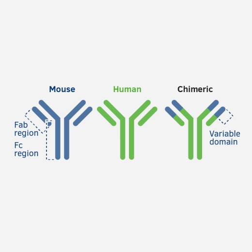
Human Chimeric AAV Antibodies
Reliable assay controls to be used when detecting AAV antibodies in human sera.